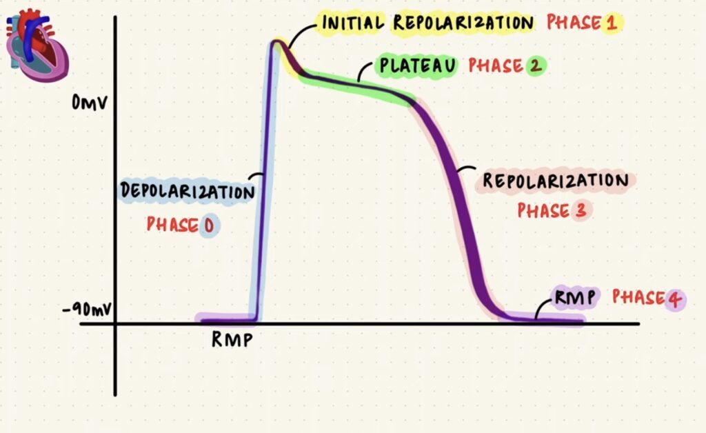
The cardiac action potential is a sequence of electrical events that occur in the heart muscle cells (myocytes) to trigger their contraction. These action potentials are essential for the proper functioning of the heart, ensuring coordinated and rhythmic contractions. The action potential in cardiac cells is typically divided into five phases (0-4).
Phases of the Cardiac Action Potential
- Phase 0: Rapid Depolarisation
- Mechanism: When the membrane potential reaches a threshold, voltage-gated sodium (Na⁺) channels open, allowing Na⁺ ions to rush into the cell.
- Result: The cell’s membrane potential becomes more positive (depolarises rapidly).
- Ion Movement: Na⁺ influx.
2. Phase 1: Initial Repolarisation
- Mechanism: Na⁺ channels close, and transient outward potassium (K⁺) channels open, causing a brief efflux of K⁺.
- Result: A slight repolarisation occurs, creating a small downward deflection after the peak of the action potential.
- Ion Movement: K⁺ efflux.
3. Phase 2: Plateau Phase
- Mechanism: Voltage-gated calcium (Ca²⁺) channels open, allowing Ca²⁺ ions to enter the cell, while K⁺ channels remain open, causing a balance between Ca²⁺ influx and K⁺ efflux.
- Result: The membrane potential remains relatively stable and prolonged, sustaining the contraction of the cardiac muscle.
- Ion Movement: Ca²⁺ influx balanced by K⁺ efflux.
4. Phase 3: Rapid Repolarisation
- Mechanism: Ca²⁺ channels close, and delayed rectifier K⁺ channels remain open, increasing K⁺ efflux.
- Result: The membrane potential returns to its resting level (repolarises).
- Ion Movement: K⁺ efflux.
5. Phase 4: Resting Membrane Potential
- Mechanism: The membrane potential is maintained by the Na⁺/K⁺ ATPase pump, which actively transports Na⁺ out of the cell and K⁺ into the cell, and by K⁺ leak channels.
- Result: The cell remains at its stable resting potential until the next depolarisation.
- Ion Movement: Na⁺/K⁺ pump activity (3 Na⁺ out, 2 K⁺ in).
Types of Cardiac Cells and Their Action Potentials
- Non-Pacemaker Cells (Contractile Cells):
- Location: Atrial and ventricular myocytes.
- Action Potential: Follows the five-phase model described above.
- Function: Responsible for the contraction of the heart muscle.
2. Pacemaker Cells (Autorhythmic Cells):
- Location: SA node, AV node.
- Action Potential Characteristics:
- Phase 4: Spontaneous depolarisation due to funny current (If) and transient calcium (T-type Ca²⁺) channels.
- Phase 0: Depolarisation due to the opening of long-lasting calcium (L-type Ca²⁺) channels.
- Phase 3: Repolarisation due to K⁺ efflux.
- Function: Generate spontaneous action potentials to set the heart rate.
Comparison of Non-Pacemaker and Pacemaker Action Potentials
| Feature | Non-Pacemaker Cells | Pacemaker Cells |
|---|---|---|
| Resting Membrane Potential | Stable, around -90 mV | Unstable, slowly depolarises |
| Phase 0 (Depolarisation) | Rapid, due to Na⁺ influx | Slow, due to Ca²⁺ influx |
| Phase 1 (Initial Repolarisation) | Present, brief K⁺ efflux | Absent |
| Phase 2 (Plateau) | Prolonged, Ca²⁺ influx and K⁺ efflux | Absent |
| Phase 3 (Repolarisation) | K⁺ efflux | K⁺ efflux |
| Phase 4 (Resting/Prepotential) | Stable, maintained by Na⁺/K⁺ pump | Unstable, gradual depolarisation due to funny current (If) |
Clinical Relevance
- Arrhythmias:
- Cause: Abnormalities in the generation or conduction of action potentials.
- Types: Bradycardia (slow heart rate), tachycardia (fast heart rate), fibrillation (irregular rhythm).
2. Pharmacological Interventions:
- Class I Antiarrhythmics: Na⁺ channel blockers, affect Phase 0.
- Class II Antiarrhythmics: Beta-blockers, affect Phase 4 and decrease heart rate.
- Class III Antiarrhythmics: K⁺ channel blockers, prolong Phase 3.
- Class IV Antiarrhythmics: Ca²⁺ channel blockers, affect Phase 2 (non-pacemaker) and Phase 0 (pacemaker).
Understanding the cardiac action potential is crucial for diagnosing and treating various heart conditions, ensuring proper electrical activity and effective contractions of the heart muscle.
Reference:
