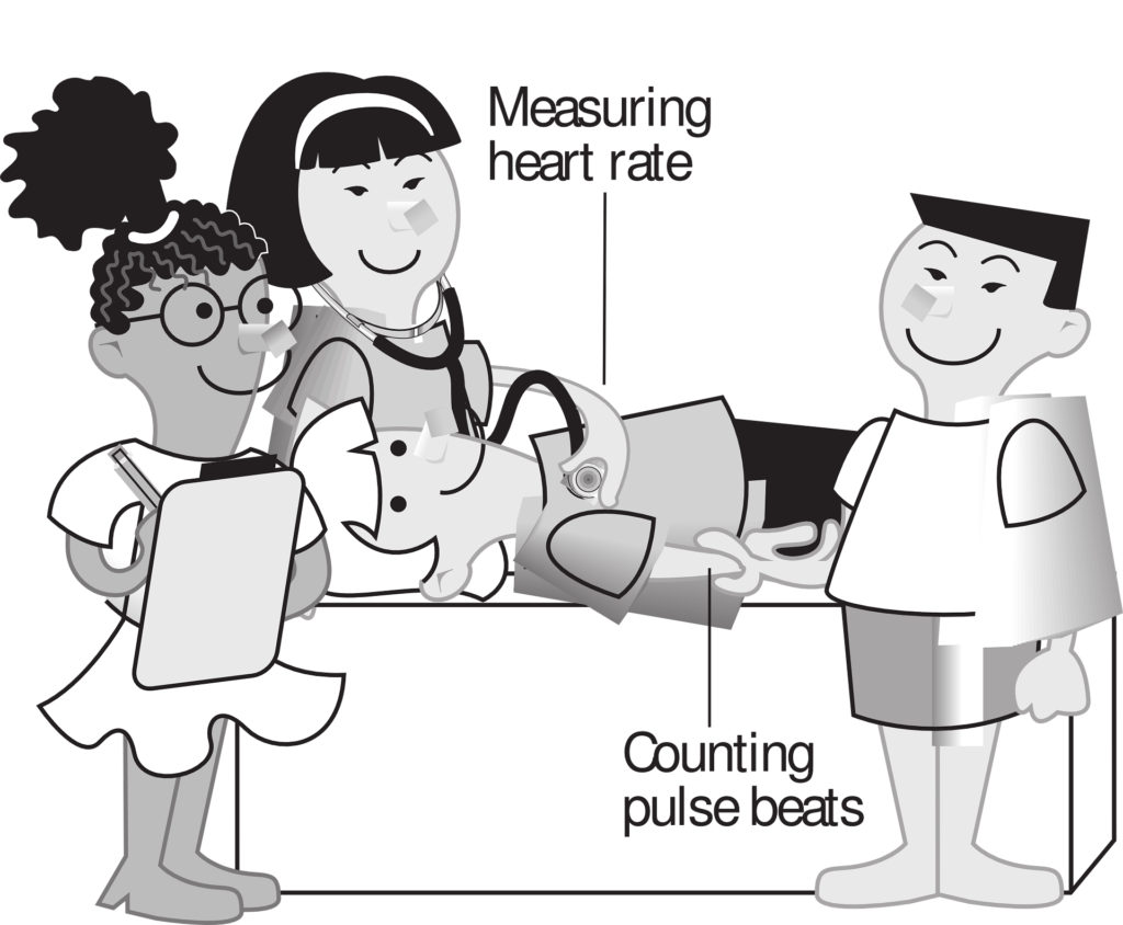
Assessing both central and peripheral pulses is crucial for evaluating a patient’s cardiovascular health. Here’s a comprehensive guide to understanding and assessing these pulses:
Central Pulses
Central pulses are closer to the heart and provide information about the heart’s direct output and major blood vessel health.
- Carotid Pulse:
- Location: Along the side of the neck, medial to the sternocleidomastoid muscle.
- Method: Use the pads of the index and middle fingers. Press gently to avoid stimulating the carotid sinus.
- Significance: Indicates central blood flow and cardiac output. A strong, regular carotid pulse suggests good heart function and blood flow. Weak or absent carotid pulses can indicate severe atherosclerosis or shock.
- Femoral Pulse:
- Location: In the groin, halfway between the pubic symphysis and the anterior superior iliac spine.
- Method: Use the pads of the index and middle fingers to palpate deeply.
- Significance: Indicates central blood flow to the lower extremities. It can be used to assess for aortic insufficiency or peripheral arterial disease. Absent femoral pulses can indicate severe vascular disease or aortic dissection.
Peripheral Pulses
Peripheral pulses are located farther from the heart and help assess blood flow to the extremities.
- Radial Pulse:
- Location: On the lateral aspect of the wrist, just proximal to the thumb.
- Method: Use the pads of the index and middle fingers to palpate the pulse.
- Significance: Easily accessible and commonly used to assess heart rate and rhythm. Weak or absent radial pulses can indicate peripheral artery disease or poor perfusion.
- Brachial Pulse:
- Location: On the medial aspect of the arm, just above the elbow.
- Method: Use the pads of the index and middle fingers to palpate.
- Significance: Important in infants and during blood pressure measurement. Weak or absent brachial pulses can indicate peripheral artery disease or blockages.
- Popliteal Pulse:
- Location: Behind the knee, in the popliteal fossa.
- Method: Use the pads of the index and middle fingers, pressing deeply into the popliteal fossa.
- Significance: Difficult to palpate; indicates blood flow to the lower leg. Absent popliteal pulses can indicate peripheral artery disease or popliteal artery occlusion.
- Posterior Tibial Pulse:
- Location: Behind the medial malleolus of the ankle.
- Method: Use the pads of the index and middle fingers to palpate.
- Significance: Assesses blood flow to the foot. Weak or absent posterior tibial pulses can indicate peripheral artery disease.
- Dorsalis Pedis Pulse:
- Location: On the dorsum of the foot, just lateral to the extensor tendon of the big toe.
- Method: Use the pads of the index and middle fingers to palpate.
- Significance: Assesses blood flow to the foot. Weak or absent dorsalis pedis pulses can indicate peripheral artery disease.
Performing the Assessment
General Steps:
- Preparation:
- Ensure the patient is relaxed and in a comfortable position.
- Explain the procedure to the patient to reduce anxiety.
- Palpation:
- Use the pads of your index and middle fingers for palpation.
- Press gently but firmly, enough to feel the pulse without occluding the vessel.
- Assess for the rate, rhythm, strength, and equality of the pulses.
- Comparison:
- Compare pulses bilaterally to check for symmetry.
- Note any differences in strength or absence of pulses, which can indicate vascular issues.
Clinical Implications
- Rate and Rhythm:
- Normal Rate: 60-100 beats per minute (bpm).
- Tachycardia: >100 bpm.
- Bradycardia: <60 bpm.
- Regular Rhythm: Evenly spaced beats.
- Irregular Rhythm: Unevenly spaced beats, seen in conditions like atrial fibrillation.
- Strength:
- Bounding Pulse: May indicate high cardiac output or hyperthyroidism.
- Weak/Thready Pulse: May indicate low cardiac output, shock, or dehydration.
- Equality:
- Symmetrical Pulses: Normal finding.
- Asymmetrical Pulses: May indicate vascular obstruction or anatomical anomalies.
Summary
Assessing central and peripheral pulses is vital in evaluating the cardiovascular status of a patient. Central pulses provide information about the heart’s output and major arterial health, while peripheral pulses help assess blood flow to the extremities. Proper technique and careful interpretation of pulse characteristics can provide valuable insights into a patient’s cardiovascular health.
