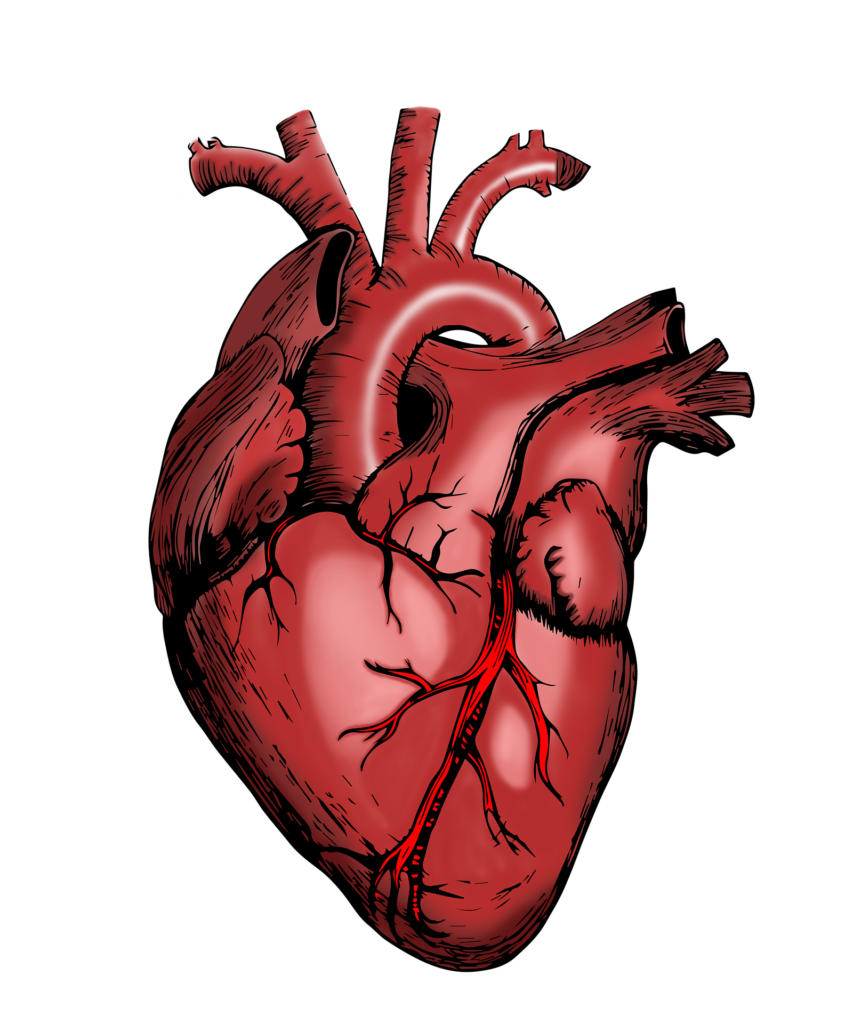
The heart’s conduction system is a network of specialised cardiac muscle cells responsible for initiating and conducting electrical impulses throughout the heart. This system ensures that the heart beats in a coordinated and efficient manner to pump blood effectively. Here is an in-depth overview of the conduction system, including its components, pathway of electrical impulses, clinical significance, and more detailed information.
Components of the Conduction System
- Sinoatrial (SA) Node:
- Location: Upper part of the right atrium near the opening of the superior vena cava.
- Function: Acts as the natural pacemaker of the heart, initiating electrical impulses that spread through the atria. These impulses cause atrial contraction.
- Rate: Generates impulses at a rate of 60-100 beats per minute under normal conditions.
- Characteristics: Contains pacemaker cells that possess automaticity, allowing them to depolarize spontaneously without external stimuli.
- Atrioventricular (AV) Node:
- Location: Lower part of the right atrium near the interatrial septum, close to the tricuspid valve.
- Function: Receives impulses from the SA node and delays them slightly to allow the ventricles to fill with blood. Then, it transmits impulses to the ventricles.
- Delay: The delay at the AV node is approximately 0.1 seconds, providing sufficient time for ventricular filling.
- Characteristics: Slow conduction velocity due to smaller fibers and fewer gap junctions, contributing to the delay.
- Bundle of His (Atrioventricular Bundle):
- Location: Runs from the AV node down the interventricular septum.
- Function: Transmits electrical impulses from the AV node to the bundle branches.
- Characteristics: Divides into the right and left bundle branches, ensuring the impulses reach both ventricles.
- Right and Left Bundle Branches:
- Location: Run along the right and left sides of the interventricular septum, respectively.
- Function: Carry impulses from the Bundle of His to the Purkinje fibers in the respective ventricles.
- Characteristics: The left bundle branch further divides into anterior and posterior fascicles, ensuring comprehensive activation of the left ventricle.
- Purkinje Fibers:
- Location: Spread throughout the ventricles, beneath the endocardium.
- Function: Rapidly conduct impulses to the ventricular muscle cells, causing coordinated ventricular contraction.
- Characteristics: Have the fastest conduction velocity in the heart, ensuring a rapid and synchronised ventricular contraction.
Pathway of Electrical Impulses
- Initiation at the SA Node:
- The SA node generates an electrical impulse.
- The impulse spreads through the atria, causing atrial depolarization and contraction (seen as the P wave on an ECG).
- Conduction to the AV Node:
- The impulse reaches the AV node, where it is delayed.
- This delay allows the ventricles to complete their filling phase.
- Transmission through the Bundle of His:
- The impulse moves from the AV node to the Bundle of His.
- Distribution via Bundle Branches:
- The impulse travels down the right and left bundle branches.
- Dissemination through Purkinje Fibers:
- The impulse spreads rapidly through the Purkinje fibers, causing ventricular depolarisation and contraction (seen as the QRS complex on an ECG).
- Repolarisation:
- After the ventricles contract, they repolarise, which is seen as the T wave on an ECG.
ECG Representation of Conduction
- P Wave: Represents atrial depolarization initiated by the SA node.
- PR Interval: Time taken for the electrical impulse to travel from the SA node through the AV node to the ventricles.
- QRS Complex: Represents ventricular depolarisation as the impulse travels through the bundle branches and Purkinje fibers.
- ST Segment: Represents the time during which the ventricles are depolarised.
- T Wave: Represents ventricular repolarisation.
Clinical Significance
- Arrhythmias:
- Atrial Fibrillation: Rapid, irregular atrial activity leading to an irregular heart rhythm.
- Atrial Flutter: Rapid but regular atrial contractions.
- Ventricular Tachycardia: Rapid ventricular contractions that can compromise cardiac output.
- Ventricular Fibrillation: Disorganised ventricular contractions, leading to cardiac arrest if untreated.
- Heart Blocks:
- First-degree Heart Block: Prolonged PR interval due to delayed conduction through the AV node.
- Second-degree Heart Block: Intermittent failure of atrial impulses to conduct to the ventricles. This can be further classified into:
- Type I (Wenckebach or Mobitz I): Progressive lengthening of the PR interval until a beat is dropped.
- Type II (Mobitz II): Sudden failure of conduction without preceding PR interval prolongation.
- Third-degree (Complete) Heart Block: No impulses conduct from the atria to the ventricles, requiring a pacemaker.
- Bundle Branch Blocks:
- Right Bundle Branch Block (RBBB): Delayed conduction through the right bundle branch, seen as a widened QRS complex on an ECG.
- Left Bundle Branch Block (LBBB): Delayed conduction through the left bundle branch, also seen as a widened QRS complex. It can affect the diagnosis of myocardial infarction.
- Accessory Pathways:
- Wolff-Parkinson-White (WPW) Syndrome: Presence of an accessory pathway (Bundle of Kent) that bypasses the AV node, leading to pre-excitation of the ventricles and a characteristic delta wave on the ECG.
Detailed Implications and Treatments
- Pacemakers: Used to treat symptomatic bradycardias or heart blocks by providing regular electrical impulses to stimulate heartbeats.
- Implantable Cardioverter-Defibrillators (ICDs): Used in patients at risk for sudden cardiac death due to ventricular tachycardia or fibrillation.
- Ablation Therapy: Used to treat arrhythmias by destroying abnormal conduction pathways or areas of ectopic focus within the heart.
- Medications: Antiarrhythmic drugs, beta-blockers, and calcium channel blockers are used to manage arrhythmias and modify conduction properties.
Summary
The heart’s conduction system is essential for maintaining a coordinated and effective heartbeat. Understanding its components and function is crucial for diagnosing and managing various cardiac conditions. By assessing and interpreting ECG changes and clinical presentations, healthcare providers can make informed decisions to ensure optimal cardiac health and patient outcomes.
