JVP – Measures central venous pressure (Right atrial pressure)
JVP is a biphasic pulse seen along the sterno-cleio-mastoid muscle of the neck.
JVP is important physical exam to evaluate cardiac conditions.
You can do this because there is no valve in the internal jugular vein
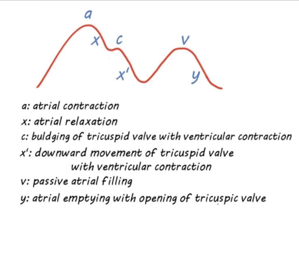
Normal JVP wave consists of 3 positive (+) waves and 2 negative (-) trough.
When the atria contracts, bloods moves from the atria to the ventricles and puts pressure on the atria. This causes the back pressure that is exerted by atria on the veins.

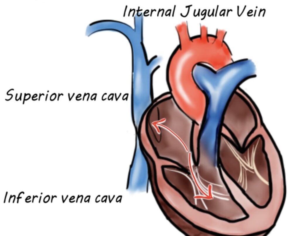
Here are IVC SVC & IJV, when there is rise in pressure in the right atria, this gets reflected in the SVC, which in turn reflected to right IJV, thus when the right atria contracts, we see a rise in the wave form giving a wave
As the atria relax the pressure drops and is reflected by X descent.
The further rise in the C wave , the tricuspid valves closes as ventricle systole occurs. This small notch represent ventricular contraction as a C wave.
As ventricle contracts, it pushes tricuspid valves. into the right atria , increasing the pressure averse slightly to give a small peak.
x’ (Xprime) corresponds with ventricular contractions, as ventricular contracts in the systole, it drags the pressure down with it causing a downward displacement of the tricuspid valves. This decreases the pressure in the right atria and we can see downward wave.
The V wave corresponds to the venous filling, when the tricuspids valves are closed, the right atria begins to fill with blood, increasing the pressure in the right atria and creating the 2nd peak or V wave. when the atria completely fills with blood, this generates enough pressure to open the tricuspids valves allowing blood to pass from right atria to right ventricles, this reflected with Y descent.
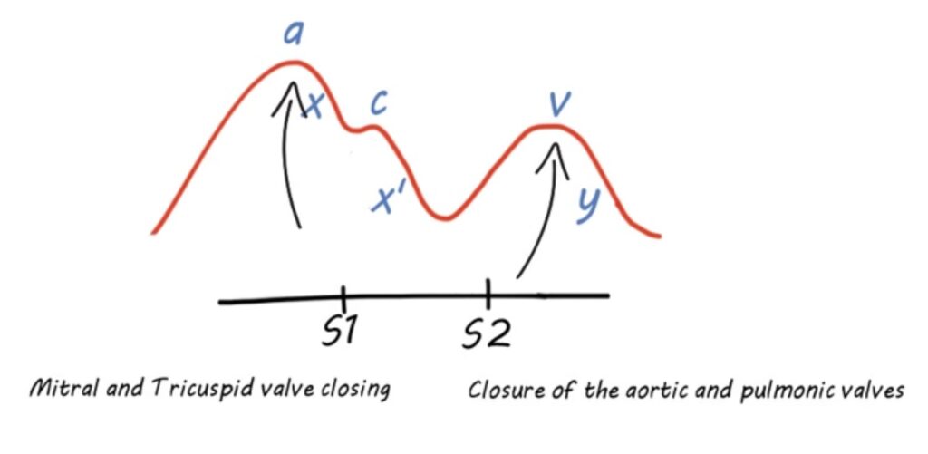
How Biphasic pulse corresponds to heart sounds ?
The first pulse is observed right before S1 ( closure of mitral and tricuspid valves)
2nd pulse is observed right after S2 ( Closure of aortic and pulmonic valves)


