Altered Physiology in Relation to Peripheral Vascular Disease (PVD)
Peripheral Vascular Disease (PVD) encompasses a range of conditions affecting the blood vessels outside of the heart and brain, including both arterial and venous components. Here’s a detailed look at the altered physiology associated with both types of PVD:
1. Arterial Peripheral Vascular Disease (PAD)
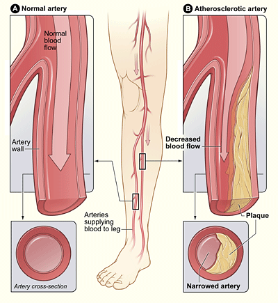
Arterial PVD primarily involves atherosclerosis, where plaque buildup narrows or blocks the arteries, reducing blood flow to the limbs.
- Atherosclerosis: Plaque formation narrows arteries, leading to decreased blood flow (ischaemia) to the affected limbs.
- Plaque Instability: Chronic inflammation can lead to unstable plaques, increasing the risk of rupture and acute arterial occlusion.
- Altered Muscle Metabolism: Ischaemia forces muscles into anaerobic metabolism, causing pain (intermittent claudication).
- Reduced Blood Flow: Decreased oxygen delivery leads to tissue ischemia, particularly during exercise.
- Endothelial Dysfunction: Impaired nitric oxide production and chronic inflammation exacerbate arterial narrowing.
- Sympathetic Nervous System Activation: Increased sympathetic tone leads to further vasoconstriction and reduced blood flow.
- Complications: Include critical limb ischaemia and acute limb ischaemia, leading to chronic pain, ulcers, and potential limb loss.
2. Venous Peripheral Vascular Disease
Venous PVD involves conditions that affect the veins, leading to impaired blood flow back to the heart.
- Venous Insufficiency: Valves in the veins may become damaged or weakened, allowing blood to flow backward (venous reflux) and pool in the legs.
- Chronic Venous Insufficiency (CVI): Persistent venous reflux leads to increased venous pressure, causing symptoms such as leg swelling, pain, and skin changes (e.g., venous stasis ulcers).
- Deep Vein Thrombosis (DVT): Blood clots may form in the deep veins of the legs, obstructing blood flow and potentially leading to pulmonary embolism if the clot dislodges and travels to the lungs.
- Varicose Veins: Enlarged, twisted veins near the surface of the skin that result from valve dysfunction and increased venous pressure.
Role of Inflammation and Lymphatic System in Arterial and Venous PVD
- Inflammation: Chronic inflammation contributes to the progression of both arterial and venous PVD by promoting plaque development in arteries and exacerbating venous wall damage and dysfunction. (Cellulitis )
- Lymphatic System: Dysfunction in the lymphatic system may exacerbate symptoms in both types of PVD. In arterial PVD, impaired lymphatic drainage may contribute to tissue oedema and inflammation, while in venous PVD, compromised lymphatic function may exacerbate venous congestion and skin changes.
Summary
Peripheral Vascular Disease encompasses both arterial (primarily PAD) and venous conditions. Arterial PVD is characterised by plaque buildup and narrowed arteries, leading to tissue ischaemia, while venous PVD involves impaired venous return and may result in symptoms such as leg swelling and venous ulcers. Inflammation and lymphatic dysfunction play roles in the pathophysiology of both types of PVD, contributing to disease progression and symptomatology. Understanding these mechanisms is crucial for effective diagnosis and management strategies tailored to each patient’s condition.
